Among several key imaging modalities available today, magnetic resonance imaging (MRI) comes forth in diagnostic applications, particularly unrivaled in neurological assessment, due to its noninvasive nature and excellent soft tissue contrast. Signals measured in MRI reflect various chemical properties of tissues including T1, T2 and PD parameters. This signal dependence on a multitude of tissue parameters allows for MRI examination of the same anatomy with multiple distinct ımage contrasts, unlike many other imaging modalities. Depending on the desired contrast among tissues, MRI acquisitions can be tailored to produce T1-weighting (e.g., for white/gray matter separation), T2-weighting (e.g., for liquid/soft tissue separation) or PD-weighting (e.g., for fat-liquid/soft tissue separation). In turn, availability of images of the same anatomical region under multiple complementary contrasts increases the sensitivity and specificity of diagnostic evaluations.
While multi-contrast MRI exams are tremendously helpful for improving diagnostic accuracy, each additional contrast adds up 6-10 minutes to the overall MRI protocol for a single subject. This excessive demand for scan time may not be permissible in the clinic due to overworked radiological equipment, and challenges can arise even with a basic set of MRI scans due to quality limitations with image artifacts. Therefore, there is a dire need to shorten MRI scan times in order to increase patient comfort, prevent image artifacts and reduce the cost of examination. These technological advances are particularly vital to enabling pervasive use of MRI in pediatric and elderly populations.
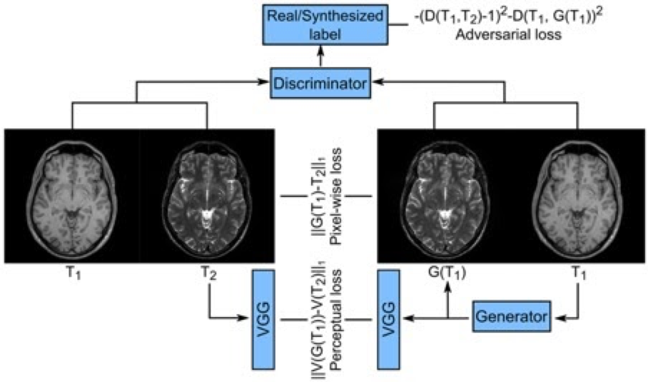
In our projects, we focus on innovative technologies that increase the quality and efficiency of MRI examinations. We are working on data-driven deep learning techniques in order to overcome the deficiencies of existing MRI methods. In this context, we are developing artificial neural network models that can successfully recover diagnostic-quality images from aggressively shortened MR exams, and that can synthesize images of missing MRI contrast subsequent to the actual exam.
The ultimate goal of our projects is to provide an advanced imaging technology that significantly improves the clinical use of MRI by enabling high scan efficiency and contrast diversity. The deep learning technology we developed will reach acceleration factors, spatial resolution, and processing time beyond existing techniques. Thus, significant improvements can be achieved in the diagnosis and treatment follow-up of many neurological and neurovascular diseases.
Sample publications:
Dar SUH, Yurt M, Ildiz ME, Shahdloo M, Tinaz B, Çukur T. Prior-Guided Image Reconstruction for Accelerated Multi-Contrast MRI via Generative Adversarial Networks IEEE Journal of Selected Topics in Signal Processing, 2020.
Dar SUH, Ozbey M, Catli AB, Çukur T. A transfer-learning approach for accelerated MRI using deep neural networks. Magnetic Resonance in Medicine. 2020 August;84(2):663–685.
Dar SUH, Yurt M, Karacan L, Erdem A, Erdem E, Çukur T. Image synthesis in multi-contrast MRI with conditional generative adversarial networks. IEEE Transaction on Medical Imaging. 2019 Oct;38(10):2375-2388.
Gozcu B, Mahabadi RK, Li YH, Ilicak E, Çukur T, Scarlett J, Cevher V. Learning-based compressive MRI. IEEE Transactions on Medical Imaging. 2018 Jun;37(6):1394-1406.